
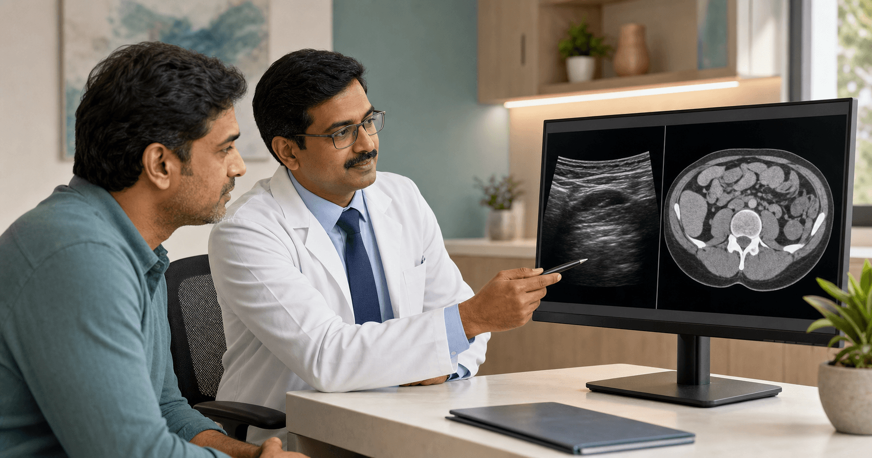
Is ultrasound or CT scan better for a suspected hernia?
Neither test is always better. Ultrasound can assess a superficial or groin swelling dynamically while you stand, cough or strain. CT gives a wider view of the abdominal wall and internal structures, which may help with large, recurrent, incisional or unclear hernias. The safest choice follows examination rather than self-booking the most expensive scan.
A hernia is tissue pushing through a weak area in the muscle or abdominal wall. A scan may show the defect, its contents and nearby anatomy, but the report still needs to be matched with symptoms and a physical examination.
Fast decision rule: if the bulge is obvious and reduces, book a routine surgical review; if symptoms repeat without a clear bulge, ask whether dynamic ultrasound or another test is useful; if the swelling is suddenly painful, stuck, discoloured or linked with vomiting, seek urgent care instead of waiting for an outpatient scan.
When can a surgeon diagnose a hernia without imaging?
A surgeon can often identify a straightforward groin, umbilical or abdominal-wall hernia from the symptom pattern and examination. The swelling may become clearer while standing, coughing or gently straining and may reduce while lying down. A bulge beside an old operation scar is covered in the /articles/incisional-hernia-symptoms-after-surgery-bhopal patient guide.
Mayo Clinic says an inguinal hernia diagnosis is usually made through physical examination, while ultrasound, CT or MRI may be ordered when the diagnosis is not readily apparent: https://www.mayoclinic.org/diseases-conditions/inguinal-hernia/diagnosis-treatment/drc-20351553.
This avoids unnecessary testing. Bring a clear standing photo if the swelling disappears before the appointment, but do not repeatedly force a painful lump back in. For a symptom-first checklist, read /articles/inguinal-hernia-symptoms-men-bhopal or /articles/hernia-symptoms-in-women-bhopal.
When may ultrasound be useful for a hernia?
Ultrasound may be useful for a small, superficial, groin or intermittent swelling, especially when the examiner wants to see what happens during coughing, straining or a change in position. It has no ionising radiation, but its usefulness depends on the body area, the operator and whether the hernia appears during the test.
The American College of Radiology rates pelvic ultrasound as usually appropriate for initial imaging of a suspected groin hernia and abdominal ultrasound as usually appropriate for a suspected abdominal-wall hernia: https://acsearch.acr.org/docs/3158169/Narrative/.
An ultrasound report should answer practical questions: where is the defect, what moves through it, does it change with strain, and is another swelling being considered? A normal scan does not automatically explain persistent pain; return to the clinician if symptoms continue.
When may a CT scan be useful for a hernia?
CT may be useful when a hernia is deep, large, recurrent, near an old operation scar, difficult to define on examination, or when the clinician needs a broader view of abdominal anatomy and possible complications. CT is not automatically required for every visible, uncomplicated bulge.
The American College of Radiology lists CT abdomen and pelvis as usually appropriate in several suspected abdominal-wall and groin-hernia scenarios. CT uses ionising radiation, and some studies use contrast, so pregnancy possibility, kidney problems, allergies and earlier scans should be discussed before the test: https://www.radiologyinfo.org/en/info/abdominct.
If the swelling has returned after an earlier repair, carry the previous operation note and mesh details when available. The focused /articles/recurrent-hernia-after-surgery-bhopal guide explains why old records can change the imaging and repair discussion.
How do ultrasound and CT compare for hernia evaluation?
The useful comparison is not simply cheaper versus more detailed. It is whether the test can answer the clinical question with the least burden. Ultrasound is dynamic and radiation-free; CT offers a wider anatomical view but involves radiation and may involve contrast.
Decision table: obvious reducible surface bulge — examination may be enough; small or intermittent groin swelling — dynamic ultrasound may help; large incisional or recurrent hernia — CT may help map anatomy; pregnancy possibility — tell the clinician before imaging; severe pain with vomiting or a stuck bulge — emergency assessment comes before routine test shopping.
No scan should be interpreted in isolation. Report wording such as fat-containing, bowel-containing, reducible, incarcerated, defect size or recurrent must be connected with current symptoms and examination.
What should you ask after receiving a hernia scan report?
Ask whether the report finding matches your symptoms, whether the hernia is reducible, whether bowel is involved, whether any urgent feature is suspected, and whether observation, planned repair or another evaluation is being discussed. Do not assume every report-labelled hernia needs immediate surgery.
Bring the report and images or disc, previous scans, operation notes, discharge summaries, medicine list, blood-thinner details, diabetes and blood-pressure records, allergy history and pregnancy status when relevant. Write down when the bulge appears and what makes it worse.
For a Bhopal consultation, Dr. Rajesh Kanungo can review the examination and reports before explaining whether no further imaging, repeat dynamic ultrasound, CT, observation, open repair or laparoscopic repair should be considered. Use /services/laparoscopic-surgery or /contact to arrange a diagnosis-first review.
Which hernia warning signs should go to emergency care?
A suddenly painful, hard or stuck swelling with vomiting, abdominal bloating, inability to pass stool or gas, fever, redness, purple or dark skin, fainting, confusion or rapid deterioration needs urgent medical assessment. Do not delay emergency care to obtain a routine ultrasound or CT appointment.
NIDDK lists sudden severe pain, a bulge that becomes larger or no longer goes back in, fever, redness, bloating, nausea and vomiting as warning signs of a stuck or strangulated inguinal hernia: https://www.niddk.nih.gov/health-information/digestive-diseases/inguinal-hernia. MedlinePlus also warns that sudden pain, nausea and vomiting can occur when a hernia becomes trapped or strangulated: https://medlineplus.gov/hernia.html.
Call the hospital or go to the nearest emergency department for severe or worsening symptoms. For a fuller emergency checklist, read /articles/strangulated-hernia-symptoms-bhopal. This article is general patient education, not a diagnosis, prescription or replacement for an in-person assessment.
Which medical sources support this guide?
This guide was cross-checked against the American College of Radiology Appropriateness Criteria for hernia imaging at https://acsearch.acr.org/docs/3158169/Narrative/, Mayo Clinic inguinal hernia diagnosis guidance at https://www.mayoclinic.org/diseases-conditions/inguinal-hernia/diagnosis-treatment/drc-20351553, RadiologyInfo CT abdomen and pelvis guidance at https://www.radiologyinfo.org/en/info/abdominct, NIDDK inguinal hernia guidance at https://www.niddk.nih.gov/health-information/digestive-diseases/inguinal-hernia, and MedlinePlus hernia information at https://medlineplus.gov/hernia.html.
The consistent patient message is conservative: examination often comes first, imaging is selected to answer a specific uncertainty, reports require clinical interpretation, and urgent symptoms should not wait for routine outpatient testing.
Related care options
More patient guides
Types of Abdominal Hernias: A Location Guide for Bhopal Patients
Abdominal hernias are named mainly by where tissue pushes through a weak area: the groin, upper thigh, navel, upper midline, abdominal wall or an old surgical scar. Location is a useful clue, but examination—not a photo or symptom list—is what separates a hernia from other lumps and guides safe treatment.
Can You Live Without a Gallbladder? What Changes After Surgery
Yes. Most people can live a normal life without a gallbladder because the liver keeps making bile. After removal, bile flows directly into the small intestine instead of being stored. Some people notice temporary digestive changes, but persistent or severe symptoms deserve medical review.
Nail Polish Before Surgery: What Should Patients Remove?
Many hospitals ask patients to remove nail polish, gel or false nails before surgery because staff may need access to a natural nail for monitoring and assessment. The safest shortcut is simple: ask the hospital in advance, follow its written instruction, and tell the team if a product or item cannot be removed safely.
Common questions
Ultrasound can help identify some superficial, groin or intermittent hernias, especially during standing, coughing or straining. The result still needs to be interpreted with symptoms and examination.
No. Many straightforward hernias can be diagnosed by examination. CT may be useful for unclear, deep, large, recurrent or incisional hernias, or when broader abdominal anatomy needs review.
Return to the clinician who examined you. A hernia may be intermittent, or another abdominal, groin, urinary, gynecologic, hip or muscle problem may need consideration. Do not diagnose the cause from one report alone.
Ultrasound does not use ionising radiation. CT does use radiation and may use contrast, but it can answer different anatomical questions. The safer choice is the test that is clinically appropriate for the specific problem.
Seek urgent care for sudden severe pain, a hard or stuck bulge, vomiting, abdominal bloating, inability to pass stool or gas, fever, skin redness or dark colour, fainting or rapid worsening.